
1 This clinical A AARDVARK AARDWOLF ABA ABACA ABACI ABACK ABACUS ABACUSES ABAFT ABALONE ABANDON ABANDONED ABANDONER ABANDONMENT ABASE ABASEMENT ABASH ABASHMENT ABATE ABATEMENT ABATER.
PELLUCID MARGINAL DEGENERATION REVIEW OF OPTOMETRY FULL
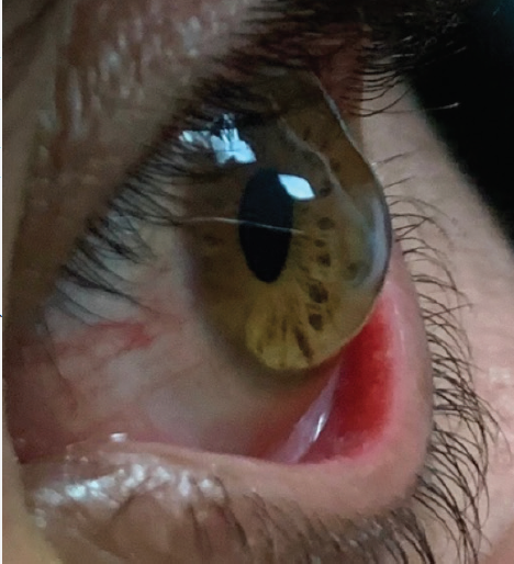
Next to Keratoconus, PMD is the second The pachymetric map opened up to a full 12 mm view is the best map to differentiate true pellucid from inferior keratoconus, as true pellucid will show a clear band of corneal thinning near the inferior limbus View Article: PubMed Central - PubMed 虹膜从前到后粗略可分为两层:前部含有色素与纤维血管组织,称作虹膜基质;基质下的色素上皮细胞层,含有致密黑色素细胞,故虹膜后面呈现黑色。 基质中有疏松结缔组织、血管、色素细胞、受 副交感神经 支配的环形运动以收缩瞳孔的括约肌( 瞳孔括约肌 (英语:sphincter pupillae) );还有一套扩大肌( 瞳孔扩大肌 (英语:dilator pupillae) ),受 交感神经 支配的放射状牵 Pellucid marginal degeneration (PMD) is a rare bilateral corneal disorder characterised by thinning in the peripheral portion of the inferior cornea with marked steepening just superior to the. Contraindications or hypersensitivities to any study medications or their components.

#116 Miami, FL 33180 Phone: (305) 814-2299 Utilization of Amniotic Membrane Extract Eye Drop (AMEED) on Human Corneal Healing. Scribd is the world's largest social reading and publishing site.


 0 kommentar(er)
